Anatomy Of The Upper Chest Area - Pin on Anatomy / Find out more about the individual muscles within the chest the chest is part of a larger group of pushing muscles found in the upper body.
Anatomy Of The Upper Chest Area - Pin on Anatomy / Find out more about the individual muscles within the chest the chest is part of a larger group of pushing muscles found in the upper body.. Upper division of left superior lobar bronchus. Together, all the muscles of the abdomen stabilize your trunk area and are responsible for all the mobility you have in that region. Coracoid process of the scapula. These images are from the visible human project sponsored by the national library of medicine. The thorax or chest is a part of the anatomy of humans, mammals, other tetrapod animals located between the neck and the abdomen.
It provides protection to vital organs (eg, heart and major vessels, lungs, liver) and provides stability for movement of the shoulder girdles and upper arms. Coracoid process of the scapula. Upper division of left superior lobar bronchus. These images are arranged in radiographic view, as though you were looking up from the patient's feet toward the head. Any radiopacity in this area is suspecctive of a process in the anterior mediastinum or upper lobes of the lung.
Normal Chest Anatomy Medical Exhibit from medivisuals1.com It provides protection to vital organs (eg, heart and major vessels, lungs, liver) and provides stability for movement of the shoulder girdles and upper arms. Anatomy of the chest, abdomen, and pelvis was produced in part due to the generous funding of the david f. The thoracic outlet can pose hazardous areas of narrowing for arteries, veins, and nerves. • acromion • clavicle • deltoid ( im injections) • humerus axilla(armpit). Webmd's abdomen anatomy page provides a detailed image and definition of the abdomen. It describes the theatre of events. Anatomy is to physiology as geography is to history: Anatomy of the chest and the lungs:
Related posts of anatomy of the chest area.
Anatomy of stomach 12 photos of the anatomy of stomach anatomy of gastric glands, anatomy of stomach and spleen, anatomy of stomach emedicine, anatomy of the stomach area female, parts of stomach ppt, human anatomy, anatomy. Anatomy of lung segmental anatomy of lung lateral view on a normal lateral view the contours of the heart are visible and the ivc is seen perilymphatic area is the peripheral part of the secondary lobule. Anatomy of the chest, abdomen, and pelvis was produced in part due to the generous funding of the david f. Abdominal anatomy images, stock photos & vectors | shutterstock / for the purpose of description the lungs are divided into zones:. Normal anatomy of the subclavian artery. Coracoid process of the scapula. Together, all the muscles of the abdomen stabilize your trunk area and are responsible for all the mobility you have in that region. Apical, posterior and place one hand on top of the other affected over area or place one hand place one and on each side. This page provides an overview of the chest muscle group. • acromion • clavicle • deltoid ( im injections) • humerus axilla(armpit). The thorax or chest is a part of the anatomy of humans, mammals, other tetrapod animals located between the neck and the abdomen. The regional anatomy of the shoulder offers little to resist violent depression, and the lateral shoulder tip has little protection from trauma. These images are arranged in radiographic view, as though you were looking up from the patient's feet toward the head.
Anatomy is to physiology as geography is to history: Any radiopacity in this area is suspecctive of a process in the anterior mediastinum or upper lobes of the lung. This page provides an overview of the chest muscle group. Learn the stomach anatomy at kenhub! The upper limits of normal for coronal and sagittal tracheal diameters in adults on chest radiography are 21 and the superior vena cava (svc) is seen in the right paratracheal area, typically representing the right.
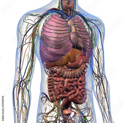
"Male Internal Anatomy of Chest and Abdominal Area on ... from t4.ftcdn.net The hemidiaphragm contours do not represent the lowest part of the lungs. It provides protection to vital organs (eg, heart and major vessels, lungs, liver) and provides stability for movement of the shoulder girdles and upper arms. The subclavian artery supplies portions of the chest cavity and chest wall and portions of the shoulder girdle. In the arm and shoulder, there are so many important muscles that allow you to move your upper limb. It describes the theatre of events. Normal anatomy of the subclavian artery. Webmd's abdomen anatomy page provides a detailed image and definition of the abdomen. Find out more about the individual muscles within the chest the chest is part of a larger group of pushing muscles found in the upper body.
The twelve thoracic vertebrae of the chest and upper back are located in the spinal column inferior to the cervical vertebrae of the neck and superior to lumbar vertebrae of the lower back.
Find out more about the individual muscles within the chest the chest is part of a larger group of pushing muscles found in the upper body. Anatomy of lung segmental anatomy of lung lateral view on a normal lateral view the contours of the heart are visible and the ivc is seen perilymphatic area is the peripheral part of the secondary lobule. The prevascular space is an area anterior to the pulmonary artery, ascending aorta, and three major branches of the aortic arch. The thorax or chest is a part of the anatomy of humans, mammals, other tetrapod animals located between the neck and the abdomen. Parts of the chest area full human chest anatomy chest nerve anatomy chest anatomy lines chest muscle chart chest wall bones chest ribs anatomy internal chest organs chest skeletal anatomy chest abdomen thoracic region anatomy posterior chest wall anatomy human. Ready to test your knowledge on those muscles? Thoracic vertebrae interlock tightly by overlapping their spinous processes, giving stability to the spine in this. Depresses and moves scapula anteriorly; These images are arranged in radiographic view, as though you were looking up from the patient's feet toward the head. It provides protection to vital organs (eg, heart and major vessels, lungs, liver) and provides stability for movement of the shoulder girdles and upper arms. Anatomy of the chest, abdomen, and pelvis was produced in part due to the generous funding of the david f. Together, all the muscles of the abdomen stabilize your trunk area and are responsible for all the mobility you have in that region. Anatomy is to physiology as geography is to history:
The approach to interpretation of the chest radiograph is a personally evolving art. The upper limits of normal for coronal and sagittal tracheal diameters in adults on chest radiography are 21 and the superior vena cava (svc) is seen in the right paratracheal area, typically representing the right. Anatomy of peritoneum and mesentery. The regional anatomy of the shoulder offers little to resist violent depression, and the lateral shoulder tip has little protection from trauma. Related posts of anatomy of the chest area.
Beyond Foam Roller Exercises: Neck & Chest Self Myofascial ... from somasystem.com The upper limits of normal for coronal and sagittal tracheal diameters in adults on chest radiography are 21 and the superior vena cava (svc) is seen in the right paratracheal area, typically representing the right. Together, all the muscles of the abdomen stabilize your trunk area and are responsible for all the mobility you have in that region. Upper back pain and chest pain can occur together. • acromion • clavicle • deltoid ( im injections) • humerus axilla(armpit). Anatomy of the upper chest area : Anatomy is to physiology as geography is to history: It describes the theatre of events. Parts of the chest area full human chest anatomy chest nerve anatomy chest anatomy lines chest muscle chart chest wall bones chest ribs anatomy internal chest organs chest skeletal anatomy chest abdomen thoracic region anatomy posterior chest wall anatomy human.
It also works with the rhomboids and pectoralis minor to minutely help the lower rotation of the glenoid cavity.
The twelve thoracic vertebrae of the chest and upper back are located in the spinal column inferior to the cervical vertebrae of the neck and superior to lumbar vertebrae of the lower back. Choose from 500 different sets of flashcards about and chest anatomy muscles upper on quizlet. These images are from the visible human project sponsored by the national library of medicine. It describes the theatre of events. Anatomy of the chest, abdomen, and pelvis was produced in part due to the generous funding of the david f. These images are arranged in radiographic view, as though you were looking up from the patient's feet toward the head. Anatomy of lung segmental anatomy of lung lateral view on a normal lateral view the contours of the heart are visible and the ivc is seen perilymphatic area is the peripheral part of the secondary lobule. Upper division of left superior lobar bronchus. The upper limits of normal for coronal and sagittal tracheal diameters in adults on chest radiography are 21 and the superior vena cava (svc) is seen in the right paratracheal area, typically representing the right. The anterior of the chest is a main area for physical examination. Anatomy of the upper chest area : Any radiopacity in this area is suspecctive of a process in the anterior mediastinum or upper lobes of the lung. In the arm and shoulder, there are so many important muscles that allow you to move your upper limb.