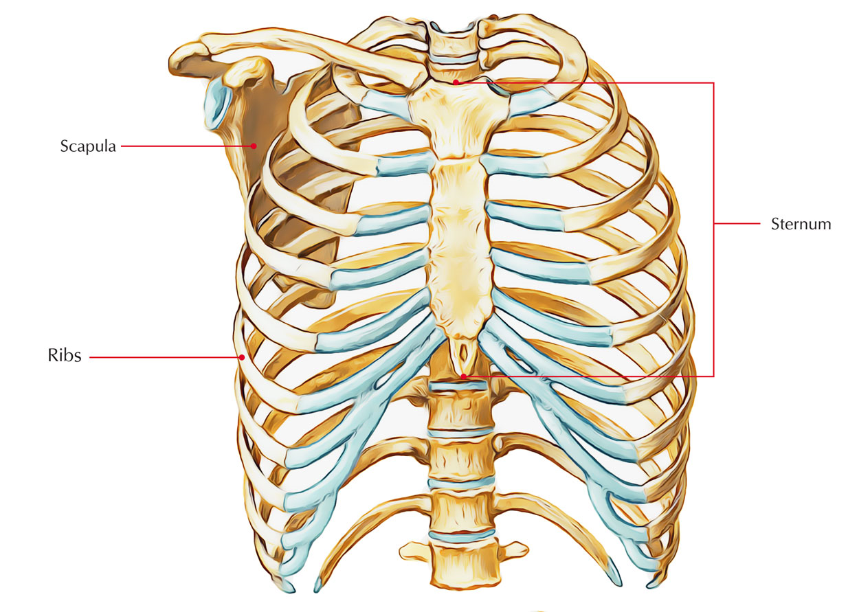
Anatomy Of Ribs And Sternum - L1 Introduction to Anatomy at University of Michigan - Ann ... : Anterior view of the thoracic cage image source.
Anatomy Of Ribs And Sternum - L1 Introduction to Anatomy at University of Michigan - Ann ... : Anterior view of the thoracic cage image source.. The classification of human ribs. Surface anatomy and surface markings bibliographic record list of illustrations subject index. Lessons on the bone markings of the ribs and sternum. Attach the ribs to the costal cartilages. True joints are generally found only at ribs 2 to 5;
Attach the ribs to the costal cartilages. Diagnose and treat somatic dysfunctions of the clavicle, sternum, and ribs. Join the sternum and clavicles. The thoracic cage consists of the 12 thoracic vertebrae, the associated intervertebral discs, 12 pairs of ribs with their costal cartilages, and the sternum. It has clear front, side, and back planes.
Ribs and sternum - Wiki1 from dornheim-anatomy.com The sternum, commonly known as the breastbone, is a long, narrow flat bone that serves as the keystone of the rib cage and stabilizes the thoracic skeleton. Learn all about this bone using our interactive anatomy image and detailed descriptions of its parts and function! Wondering what the sternum is? True ribs, false ribs, and floating ribs. There are 7 pairs or 14 total true ribs. Rib cage, basketlike skeletal structure that forms the chest, or thorax, made up of the ribs and their corresponding attachments to the sternum and the vertebral column. Ribs are not merely armour for the organs inside our torsos, as we reveal here… rib pairs one through seven are called 'true ribs' because they attach directly to the sternum. True ribs, false ribs, floating ribs.
True ribs, false ribs, and floating ribs.
True joints are generally found only at ribs 2 to 5; True ribs, false ribs, floating ribs. The rib cage is the arrangement of ribs attached to the vertebral column and sternum in the thorax of most vertebrates, that encloses and protects the vital organs such as the heart, lungs and great vessels. The sternum, commonly known as the breastbone, is a long, narrow flat bone that serves as the keystone of the rib cage and stabilizes the thoracic skeleton. The length of ribs increases from 1st to 7th rib and after that slowly falls; The thoracic cage (rib cage) is the skeletal framework of the thoracic wall, which encloses the thoracic cavity. Learn more about the skeletal system with quizzes and labelling exercises. Learn all about this bone using our interactive anatomy image and detailed descriptions of its parts and function! Sternum is a part of the skeletal system. At their anterior ends, they differ as to how they. They increase in length, curvature and amount of cartilage craniocaudally. Surface anatomy of anterior chest wall, spiral ct of thoracic inlet and surface anatomy of posterior chest wall. Ribs 1, 6, and 7 attach to the sternum by synchondroses.
This packet goes over the gross anatomy of the sternum and rib cage. Ribs eight to ten are the false ribs and are connected to the sternum indirectly via the cartilage of the rib above them. Costae are arranged in pairs and articulate with two successive vertebrae. Choose from 500 different sets of flashcards about anatomy sternum ribs on quizlet. It connects to the ribs via cartilage and forms the front of the rib cage, thus helping to protect the heart, lungs, and major blood vessels from injury.
Sternum Anatomy - Human Anatomy from www.earthslab.com Ribs 1, 6, and 7 attach to the sternum by synchondroses. Ossification.—the sternum originally consists of two cartilaginous bars, situated one on either side of the median plane and connected with the cartilages of the upper nine ribs of its. There are two classifications of ribs. Therefore in an anatomical position, the posterior end is higher and nearer the median plane in relation to the anterior end. The costotransverse ligaments in human: They increase in length, curvature and amount of cartilage craniocaudally. 5.7 sternocostal joints anterior view with right half of sternum sectioned frontally. Surface anatomy and surface markings bibliographic record list of illustrations subject index.
The chest wall is formed from the sternum anteriorly, 12 pairs of ribs, costal cartilages and intercostal muscles laterally, and the thoracic vertebrae posteriorly.
Ossification.—the sternum originally consists of two cartilaginous bars, situated one on either side of the median plane and connected with the cartilages of the upper nine ribs of its. Describe the motion of the clavicle during upper limb movement and the sternum during respiration. At their anterior ends, they differ as to how they. Each pair articulates with a different thoracic vertebra on the posterior side of the body. The thoracic cage (rib cage) is the skeletal framework of the thoracic wall, which encloses the thoracic cavity. The front plane is composed of the sternum and. All ribs articulate posteriorly with a corresponding thoracic vertebra. Rib pairs eight through ten attach indirectly through other cartilage structures, so they're. The sternum, commonly known as the breastbone, is a long, narrow flat bone that serves as the keystone of the rib cage and stabilizes the thoracic skeleton. Costae are arranged in pairs and articulate with two successive vertebrae. Learn all about this bone using our interactive anatomy image and detailed descriptions of its parts and function! Ribs 1, 6, and 7 attach to the sternum by synchondroses. Anterior view of the thoracic cage image source.
Describe the bony and cartilaginous articulations of the sternum and clavicle. The length of ribs increases from 1st to 7th rib and after that slowly falls; It lies on the anterior thoracic wall in the middle. Ribs are not merely armour for the organs inside our torsos, as we reveal here… rib pairs one through seven are called 'true ribs' because they attach directly to the sternum. The sternum, commonly known as the breastbone, is a long, narrow flat bone that serves as the keystone of the rib cage and stabilizes the thoracic skeleton.
Thorax Chest Labeled Rib Cage Sternum Photographic Print ... from imgc.allpostersimages.com It lies on the anterior thoracic wall in the middle. The number is the same in both males and females. Anatomical variations of the sternum include varying sizes of the sternal angle. At their anterior ends, they differ as to how they. Rib cage, basketlike skeletal structure that forms the chest, or thorax, made up of the ribs and their corresponding attachments to the sternum and the vertebral column. There are twelve pairs of ribs. Ribs 1, 6, and 7 attach to the sternum by synchondroses. The chest wall is formed from the sternum anteriorly, 12 pairs of ribs, costal cartilages and intercostal muscles laterally, and the thoracic vertebrae posteriorly.
Anterior view of the thoracic cage image source.
Moore'sanatomy and osteology human anatomy vertebra bones of the upper limb how to classified ribs and their shapes and fully describe human anatomy of upper parts. Try to be as accurate as you can with them. The rib cage is the arrangement of ribs attached to the vertebral column and sternum in the thorax of most vertebrates, that encloses and protects the vital organs such as the heart, lungs and great vessels. Costae are arranged in pairs and articulate with two successive vertebrae. The body is the middle part of the sternum and is also the longest. Wondering what the sternum is? The sternum or breastbone is a long flat bone located in the central part of the chest. Interactive tutorials about the ribs and sternum bones, with labeled images and diagrams featuring the beautiful illustrations of getbodysmart. The thoracic cage includes the thoracic vertebrae, sternum, ribs, and costal cartilage (see figure 1). The costotransverse ligaments in human: Regional vertebrae (cervical, thoracic, lumbar), rib, sternum, os coxae, clavicle, scapula, humerus, ulna and radius for dr. True ribs, false ribs, and floating ribs. The chest wall is formed from the sternum anteriorly, 12 pairs of ribs, costal cartilages and intercostal muscles laterally, and the thoracic vertebrae posteriorly.
The thoracic cage (rib cage) is the skeletal framework of the thoracic wall, which encloses the thoracic cavity anatomy of ribs. Sternum is a part of the skeletal system.